| The
Breasts: Some Morphological Aspects Adapted excerpt from "Os Órgãos Sexuais Femininos" Nelson Soucasaux |
 |
As it is well
known, the basic structures that, in women, give rise to the breasts,
already exist in both sexes since embryonic life: they consist of the
nipple, the areola and a rudimentary system of very small branching tubules
that originate in the nipple and spread into the subareolar connective
tissue. This branching system of tubules is formed by 15 to 20 main ducts
( canals ).
By the time of the puberal growth of the breasts, the proliferation of
each one of these ducts results in the formation of an isolated glandular
structure that corresponds to a mammary lobe. Ham observes that each breast
is constituted by the reunion of many individualized glands, each one
of them possessing an independent excretory canal through which its secretion
is conducted towards the nipple ( Ham, A.W. -"Histologia"
- Guanabara Koogan, Rio de Janeiro, 1967 ). Each mammary lobe, in turn,
consists of a great number of lobules, and each one of these lobules by
10 to 100 acini grouped together around a small collecting duct ( Haagensen,
C.D. - "Diseases of the Breast" - Saunders, Philadelphia,
London, 1956 ).
Just like all the other woman's sexual organs, both the development and
the trophicity of the breasts depend on the estrogens. Thus, the puberal
growth of the breasts is mostly due to the estrogen stimulation. There
is also a small complementary action on the part of progesterone as the
biphasic (ovulatory) cycles are established.
The puberal
development of the breasts is characterized by: 1) great proliferation
of the ductal system ( the branching canalicular structures of the breasts
) and of the connective and adipose tissue; 2) beginning of the development
of the acinar or alveolar system ( the mammary secretory structures );
3) increase in size and pigmentation of the nipples and areolas. All these
changes make the girl's originally "flat breasts" acquire the
features of those of the adult woman.
 |
 |
The estrogenic action on the breasts mostly stimulates the growth and
proliferation of the ductal ( canalicular ) system, the loose and fibrous
connective tissue that surrounds the ducts and lobules, as well as the
deposition of fat. Progesterone, besides reducing some excessive proliferative
effects of the estrogens, acting conjointly with the latter, seems to
be the main hormonal factor responsible for the development of the mammary
secretory acini ( the alveolar structures of the breasts ). There are
reports that the entire development of the lobular-alveolar system is
only completed during pregnancy.
The gestational hypertrophy of
the breasts is basically a result of the heightened levels of estrogens
and progesterone that characterize this event of women's lives. The heightened
prolactin levels that are equally typical of pregnancy possibly also contribute
to this gestational enlargement of the breasts. Though the main actions
of prolactin on the breasts consist of preparing these organs for lactation
and stimulating milk secretion, there seems to exist a synergistic action
between this pituitary hormone, the estrogens and progesterone on the puberal
and gestational development of the mammary glandular structures.
Here we must observe that, since associated with the estrogens, progesterone
and prolactin, other hormones characterized by general metabolic actions
also seem to exert a non-specific and complementary action on the mammary
development and physiology. Among them we can mention the growth hormone,
thyroxine, triiodothyronine and cortisol.
During the puberal development
of the breasts, the ducts proliferate according to a typical, increasingly
branching pattern ( like the growth of the branches in a tree ) and, at
the final extremities of their smaller branches, epithelial sprouts that
will originate the mammary acini are formed. As already stated, this great
proliferation of the mammary ducts or tubules is fundamentally due to the
estrogenic action. The development of the acinar system ( the mammary secretory
structures ) only seems to begin with the establishment of the ovulatory
cycles and the consequent appearance of progesterone along the second phase
of the cycles. Even so, the entire development of the acinar system takes
place only during pregnancy.
Also as a result of the estrogenic
action and simultaneously with the remarkable branching growth of the ductal
system, there is a great proliferation of periductal, intralobular, interlobular
and interlobar connective tissue, as well as the deposition of fat. Thus,
according to what is being exposed, we can verify that the inner structure
of the breasts is basically formed by an epithelial parenchyma and a connective
and adipose stroma.
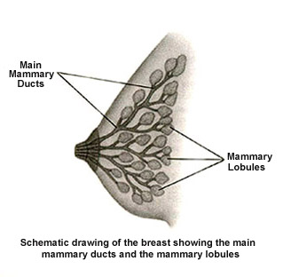
As already
mentioned, the mammary parenchyma is formed by an intricate and complex
branching system of ducts and glandular acini. In it, the diverse groupings
of acini with their respective ducts and surrounding connective tissue
constitute the mammary lobules and lobes. According to Netter's description,
the mammary ducts ". . . extend radially from the nipple toward the
chest wall, and from them sprout variable numbers of secondary tubules.
These end in epithelial masses forming the lobules or acinar structures
of the breast" ( Netter, F.H. - "The Ciba Collection of Medical
Illustrations, Vol. 2, Reproductive System" - U.S.A., 1954 ).
The mammary secretory acini are surrounded by the myoepithelial cells, capable
of contracting under the action of oxytocin. Thus, the release of oxytocin
that takes place during the act of breast feeding causes the contraction
of these myoepithelial cells and the consequent ejection of milk.
Both the quantity and the dimensions
of the ductal, acinar and lobular structures vary greatly from woman to
woman. Great variations are also found in the same woman not only according
to the phases of life and their respective hormonal influences, but also
even from one breast to the other. Almost always the major concentration
of glandular structures occupies the upper-external quadrants of the breasts.
For that reason, at clinical examination the density and consistency of
the mammary tissues is usually greater at the upper-external quadrants than
at the other ones.
As we have
seen, the mammary stroma, in the middle of which the ducts and acini are
located, consists of a mixture of fibrous and loose connective and fatty
tissue. The loose and fibrous connective tissue predominates at the parts
of the breasts in which the amount of ductal and acinar ( glandular )
structures is greater, while the fatty tissue predominates at the parts
possessing less glandular structures. As to the importance of the fibrous
and fatty tissue that constitute the mammary stroma on the degree of firmness
or flaccidity of the breasts, Netter observes that ". . . in the
absence of pregnancy and lactation, the relative amounts of fatty and
fibrous tissue determine the size and consistency of the breast".
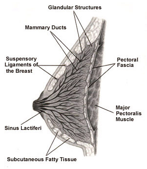
The suspensory ligaments of the
breasts constitute another group of structures of the mammary stroma that
also play an important role in the inner architecture of these organs. They
consist of thin layers of dense fibrous tissue that "encase" and
"envelop" the mammary lobes and lobules and indirectly attach
them both to the breast subcutaneous tissue and to the deep pectoral fascia.
( The deep pectoral fascia is located behind the breasts, covering the great
pectoral muscles ). Thus, the suspensory ligaments of the breasts are responsible
for the somewhat difficult anatomic task of "holding up" the entire
breast structure in its position by keeping it "anchored" both
on the skin and on the great pectoral muscle.
As to the fatty tissue that is
part of the mammary stroma, most of it is located at the subcutaneous region,
between the skin and most of the glandular structure. A thinner layer of
this tissue is found at the retromammary region, above the aforementioned
pectoral fascia. Small amounts of fatty tissue are also present in the middle
of the connective tissue that surrounds the ductal and acinar structures.
The nipples are formed mostly by dense connective tissue and smooth muscle
fibers and each one of them is internally crossed by the 15 to 20 main mammary
ducts that open up on its surface. The smooth muscle fibers surround the
mammary ducts, and their contraction cause the erection of the nipples.
The nipples also contain nerve endings especially sensitive to pressure,
whose stimulation triggers a neuroendocrine reflex responsible both for
the hypothalamic release of oxytocin and for the pituitary momentary outputs
of increased amounts of prolactin. ( These neuroendocrine events, triggered
by sucking the nipples, are fundamental during breast feeding because the
release of oxytocin cause the ejection of milk, and the pituitary "peaks"
of prolactin maintain the lacteal production. ) The nerve endings of the
nipples are also responsible for their enormous sensitivity to sexual stimulation.
The areolas also consist of dense
connective tissue containing many smooth muscle fibers. They also contain
several modified sebaceous glands that can clearly be seen on their surface.
At the subareolar region and just below the nipple the mammary ducts exhibit
short small dilations called sinus lactiferi.
Asymmetries and differences between one breast and the other in the same
woman are very frequent. They include differences not only regarding size
and shape, but also the distribution, density and relative amounts of
the several elements that constitute the mammary parenchyma and stroma.
These differences existing from one breast to the other in the same woman
are due to variations on the responsivity of the mammary tissues of each
breast to the hormonal stimulations.
![]()
Nelson Soucasaux is a gynecologist
dedicated to Clinical, Preventive and Psychosomatic Gynecology. Graduated
in 1974 by Faculdade de Medicina da Universidade Federal do Rio de Janeiro,
he is the author of several articles published in medical journals and of
the books "Novas Perspectivas em Ginecologia"
("New Perspectives in Gynecology") and "Os
Órgãos Sexuais Femininos: Forma, Função, Símbolo
e Arquétipo" ("The Female Sexual Organs: Shape, Function,
Symbol and Archetype"), published by Imago Editora, Rio
de Janeiro, 1990, 1993.
![]()
[ Home
] [ Consultório (Medical Office)
] [ Obras Publicadas (Published Works)
]
[ Novas Perspectivas em Ginecologia (New Perspectives
in Gynecology) ]
[ Órgãos Sexuais Femininos (The
Female Sexual Organs) ]
[ Temas Polêmicos (Polemical Subjects)
] [ Tópicos Diversos (Other
Topics) (Part 1) ]
[ Tópicos Diversos (Other
Topics) (Part 2 ) ] [ Tópicos
Diversos (Other Topics) (Part 3) ]
[ Ilustrações (Illustrations)
]
![]()